Vein Lump or Birthmark Since Childhood: Vascular Malformation or Varicose Vein?
✔ Medically reviewed by Dr. Ashutosh Shah - M.B., M.S., M.Ch., D.N.B., Board-Certified Plastic & Cosmetic Surgeon, Director of Elegance Clinic, Surat (22+ years’ experience).
A soft bluish lump or vein-like birthmark present since childhood, which swells when the limb hangs down or during puberty or pregnancy, is usually a venous malformation rather than varicose veins. It is diagnosed with Doppler ultrasound and MRI, and treated with compression, sclerotherapy, laser or surgery by a team, because stripping as for varicose veins can make it worse.
A blue vein lump on the arm, leg or another body area can be confusing. Some people notice it during childhood, while others become aware of it only when the affected area grows or begins to swell.
Patients often assume that any enlarged vein must be a varicose vein. However, a congenital vascular malformation behaves differently from ordinary varicose veins.
Vascular malformation treatment depends on the type of abnormal blood vessels, their depth, blood-flow characteristics and associated symptoms.
Doctors may use Doppler ultrasound, MRI and other investigations to identify the condition before recommending compression, sclerotherapy, laser treatment or surgery.
This guide explains how to recognise the differences, which warning signs require medical attention and why accurate classification matters before treatment.
Key Takeaways
-
A bluish lump present since childhood may indicate a venous malformation.
-
Varicose veins usually develop because of superficial venous valve dysfunction.
-
Venous malformations are congenital, although they may become noticeable later.
-
Puberty, pregnancy, injury and hormonal changes may influence the appearance of certain malformations.
-
Doppler ultrasound helps evaluate blood flow, while MRI shows deeper tissue involvement.
-
Treatment may include observation, compression, sclerotherapy, laser or selected surgery.
-
Standard varicose-vein stripping is not automatically suitable for a venous malformation.
-
Sudden severe pain, bleeding, rapid swelling or skin breakdown requires prompt medical assessment.
How Is a Vascular Malformation Different From Varicose Veins?
A vascular malformation is an abnormal arrangement or development of blood vessels.
These abnormalities are generally present from birth, although some become visible only later in life.
They may involve veins, arteries, capillaries, lymphatic vessels or combinations of vessel types.
Varicose veins are different.
They develop when superficial veins become enlarged and twisted, commonly because their valves do not function properly.
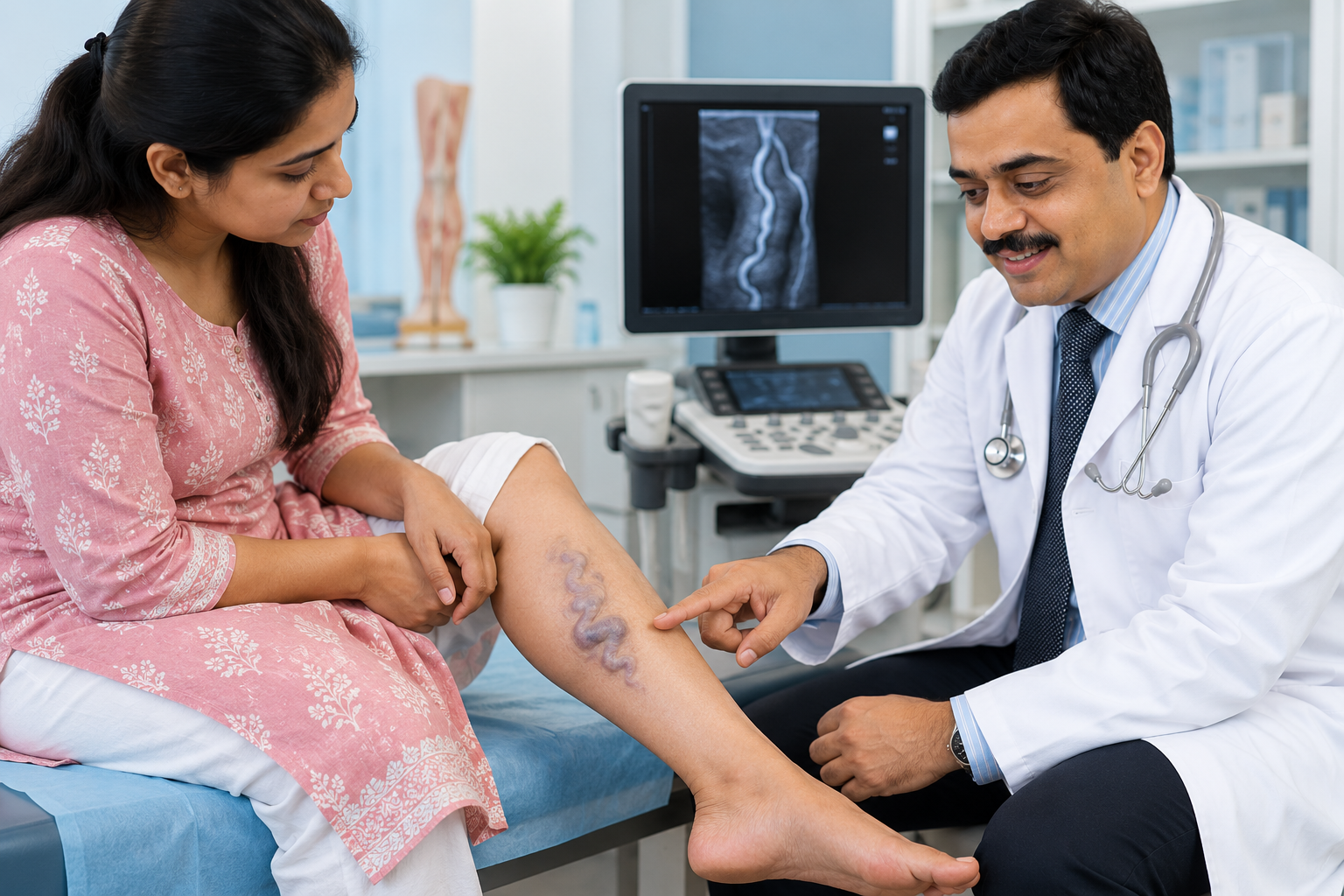
What Does a Venous Malformation Look Like?
A venous malformation may appear as:
-
A soft bluish swelling.
-
A compressible lump beneath the skin.
-
A cluster of abnormal veins.
-
A swelling that becomes fuller when the limb hangs downward.
-
A lesion that changes size with body position.
Some venous malformations are superficial, while others extend into muscles or deeper tissues.
What Do Varicose Veins Look Like?
Varicose veins often appear as enlarged, twisting veins beneath the skin.
They commonly affect the legs and may cause:
-
Aching.
-
Heaviness.
-
Swelling.
-
Itching.
-
Skin discolouration.
-
Discomfort after prolonged standing.
A vascular malformation may resemble varicose veins, but its origin and treatment requirements are different.
For further information about venous disease, see Varicose and vascular surgery.
Vascular Malformation, Varicose Vein, AVM or Haemangioma: Comparison Table
The International Society for the Study of Vascular Anomalies (ISSVA) separates vascular anomalies into vascular tumours and vascular malformations.
This classification helps clinicians avoid treating different conditions as though they were the same.
| Feature | Varicose vein | Venous malformation | Arteriovenous malformation (AVM) | Infantile haemangioma |
|---|---|---|---|---|
| Typical onset | Usually develops later | Present from birth, sometimes recognised later | Usually congenital, may become apparent later | Usually appears during early infancy |
| Appearance | Enlarged, twisted superficial veins | Soft bluish or compressible swelling | May appear warm, red or pulsating | Red or bluish vascular growth |
| Blood flow | Venous flow, often with reflux | Usually slow-flow | High-flow | Vascular tumour with characteristic growth pattern |
| Changes over time | May worsen with age or venous pressure | Often grows proportionately; may enlarge with hormonal changes | May progress or expand | Often grows in infancy, then gradually involutes |
| First imaging test | Duplex venous ultrasound | Doppler ultrasound | Doppler ultrasound | Usually clinical diagnosis; ultrasound if uncertain |
| Further imaging | Selected cases | MRI to assess extent | MRI/MRA or angiographic assessment when indicated | Imaging for deep, complex or atypical lesions |
| Common treatment | Compression, ablation or selected procedures | Compression, sclerotherapy, selected surgery | Specialist embolisation and/or surgery | Observation or medical treatment when indicated |
A haemangioma is not simply another name for a venous malformation.
Likewise, a high-flow AVM must not be treated as though it were an ordinary slow-flow venous lesion.
Why Do Vein Lumps Grow During Puberty or Pregnancy?
Some vascular malformations become more noticeable as the body grows.
A lesion that was barely visible during childhood may become larger or more symptomatic during adolescence or adulthood.
Hormonal Changes
Puberty and pregnancy involve hormonal and circulatory changes.
These changes may increase the size or visibility of certain venous malformations.
However, not every lesion behaves in the same way.
Increased Venous Pressure
A venous malformation may become fuller when the affected limb hangs downward.
This occurs because blood collects within the abnormal venous spaces.
Elevation may reduce the swelling in some patients.
Injury or Localised Clotting
Trauma, inflammation or localised clot formation within a venous malformation may cause pain and swelling.
Small calcified clots, called phleboliths, can develop in some lesions.
These may be visible on imaging.
Does a Growing Lump Mean Cancer?
Growth alone does not mean a lesion is cancerous.
Most venous malformations are not malignant tumours.
However, a rapidly changing, firm, painful or otherwise unusual lump should be assessed to confirm the diagnosis.
Which Scans Confirm the Type of Vascular Malformation?
Accurate diagnosis is essential before starting vascular malformation treatment.
The doctor first reviews when the lesion appeared, how it has changed and whether it causes pain, swelling, bleeding or functional problems.
Physical examination may assess colour, compressibility, warmth, pulsation and changes with position.
1. Doppler Ultrasound
Doppler ultrasound is often the first imaging test.
It helps assess:
-
Whether the lesion contains blood vessels.
-
The pattern and speed of blood flow.
-
Whether the lesion is slow-flow or high-flow.
-
Superficial tissue involvement.
-
Associated venous abnormalities.
A typical venous malformation is slow-flow, although very slow flow can sometimes be difficult to detect.
2. MRI
MRI is particularly useful for larger, deeper or complex vascular malformations.
It can show:
-
The size and extent of the lesion.
-
Involvement of muscles and other tissues.
-
Relationships with nearby structures.
-
Whether the malformation extends beyond the visible swelling.
MRI findings help the treating team decide whether sclerotherapy, surgery or another approach is appropriate.
3. Additional Imaging
Selected high-flow lesions may require magnetic resonance angiography or catheter angiography for detailed treatment planning.
Not every patient requires these investigations.
The imaging approach depends on the suspected vascular anomaly.
Compression, Sclerotherapy, Laser or Surgery: Which Treatment Is Suitable?
The appropriate treatment depends on the type of malformation, symptoms, location and involvement of surrounding tissues.
Not every vascular malformation requires immediate intervention.
Small, stable and asymptomatic lesions may be monitored.
Compression Therapy
Compression garments may help selected patients with venous malformations affecting the limbs.
They can reduce discomfort or swelling in some cases.
Compression does not remove the abnormal vessels.
It should be prescribed appropriately, particularly when arterial disease or other circulation problems are present.
Sclerotherapy
Sclerotherapy involves injecting a specialist medication into abnormal vascular spaces to damage their lining and encourage the treated spaces to shrink.
Image-guided sclerotherapy is commonly used for suitable venous malformations.
Possible risks include:
-
Pain and swelling.
-
Skin injury.
-
Infection.
-
Nerve irritation or injury.
-
Blood clots.
-
Damage to nearby tissues.
Several sessions may be necessary.
The procedure should be performed by clinicians experienced in treating vascular anomalies.
Laser Treatment
Laser treatment may help selected superficial vascular lesions.
The laser type and treatment depth depend on the condition.
Deep venous malformations usually cannot be adequately treated with surface laser therapy alone.
For information about appropriate superficial lesions, visit Laser for vascular lesions.
Surgery
Surgery may be considered for selected localised lesions or when a malformation causes significant pain, functional impairment or deformity.
However, extensive lesions can involve muscles, nerves and important blood vessels.
Complete removal may not be possible or advisable.
Surgery may also be combined with sclerotherapy or other treatments.
Why Standard Varicose-Vein Stripping May Be Inappropriate
A venous malformation is not simply a defective superficial vein.
It may consist of multiple abnormal vascular spaces extending through different tissue layers.
Treating it with ordinary varicose-vein stripping without proper imaging may cause bleeding, tissue injury or incomplete treatment.
A suspected venous malformation should be classified before any vein-removal procedure.
For comparison, read How varicose veins are treated.
Can a Vascular Malformation Be Removed Completely?
Some small, well-defined lesions can be removed completely.
Others are extensive or involve important surrounding structures.
In those cases, treatment may aim to:
-
Reduce pain.
-
Control swelling.
-
Improve movement.
-
Reduce bleeding.
-
Improve appearance.
-
Prevent or manage complications.
Recurrence or residual disease is possible, particularly with diffuse malformations.
Patients should understand that improvement and complete cure are not always the same.
When Does a Vein Lump Need Urgent Medical Attention?
A long-standing vascular lesion should be reassessed if its behaviour changes.
Warning signs include:
-
Sudden or severe pain.
-
Rapid swelling.
-
Significant bleeding.
-
Skin breakdown or ulceration.
-
New weakness or numbness.
-
Marked warmth or pulsation.
-
A rapidly enlarging mass.
Active bleeding that does not stop with firm direct pressure, severe limb pain with circulation changes or sudden swelling accompanied by breathing difficulty requires emergency care.
Pain can sometimes result from localised clotting within a venous malformation, but other causes must also be considered.
Patients should not attempt to puncture, drain or massage an unexplained vascular lump.
Vascular Malformation Treatment in Indore: When Should You Consult a Specialist?
A blue vein-like birthmark present since childhood should be assessed when it becomes painful, grows, affects movement or creates concerns about its appearance.
Dr. Ashutosh Shah, Plastic and Reconstructive Surgeon at Elegance Clinic, may participate in the evaluation and surgical management of suitable vascular lesions, with imaging and specialist input as required.
The care team for a complex vascular malformation may include:
-
A plastic or reconstructive surgeon.
-
An interventional radiologist.
-
A vascular specialist.
-
A dermatologist.
-
Other specialists depending on the lesion and associated conditions.
A consultation should establish the likely diagnosis and determine whether Doppler ultrasound, MRI or further specialist evaluation is required.
The treatment plan should follow the imaging findings rather than the visible appearance alone.
Can a Vascular Birthmark Be Treated for Cosmetic Reasons?
Some vascular lesions cause little physical discomfort but remain cosmetically noticeable.
Treatment may be considered when the patient understands the likely benefits, limitations and risks.
However, a cosmetic concern should not lead to treatment without first confirming the lesion type.
For example, a superficial capillary birthmark may respond to certain lasers, while a deeper venous malformation may require a completely different approach.
The Birthmark removal service page explains additional treatment considerations.
The goal is to choose a procedure appropriate for the underlying condition, not simply to remove visible colour.
Conclusion
A bluish lump or vein-like birthmark present since childhood is not necessarily a varicose vein.
Venous malformations, arteriovenous malformations, haemangiomas and ordinary varicose veins differ in their origin, blood flow and treatment requirements.
Doppler ultrasound and MRI help identify the type and extent of a lesion.
Vascular malformation treatment may involve observation, compression, sclerotherapy, laser or selected surgery, depending on the diagnosis.
The most important step is to identify the vascular anomaly correctly before attempting treatment.
A diagnosis-first approach can reduce unnecessary procedures and help patients choose a safer, more appropriate plan.
Frequently Asked Questions
Is a Vascular Malformation a Tumour?
Can a Vascular Malformation Be Removed Completely?
Why Does My Vein Lump Hurt in the Morning?
Is Sclerotherapy Safe for Children?
Can a Vascular Malformation Bleed?
Which Doctor Treats Vascular Malformations?
About the Author